桡骨骨折(肘关节外髁骨折)
来自:双语学影像;本期病例选自AuntMinnie.com
Our appreciation is extended to Dr. Manish L. Jani,Indiana University Department of Radiology,for contributing this case.

History and Radiographs
History
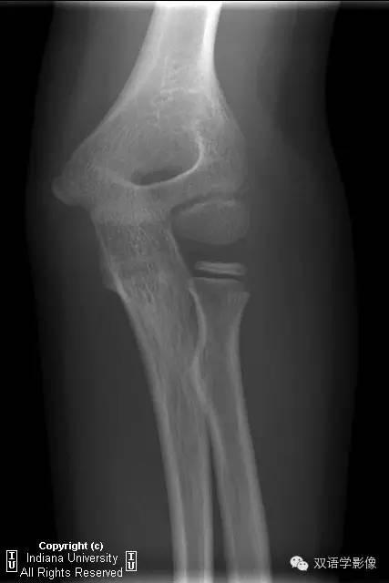
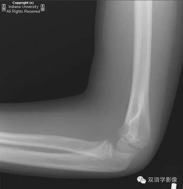
A 6-year-old girl with pain after a fall.
6岁女孩,摔伤后疼痛。
1.Which choice best characterizes the salient abnormality?
Radial head dislocation 桡骨小头脱位
Epicondylar fracture 肱骨髁上骨折
Medial condyle fracture 肱骨内髁骨折
Lateral condyle fracture 肱骨外髁骨折
Normal variant (no acute abnormality)正常变异
Additional Images
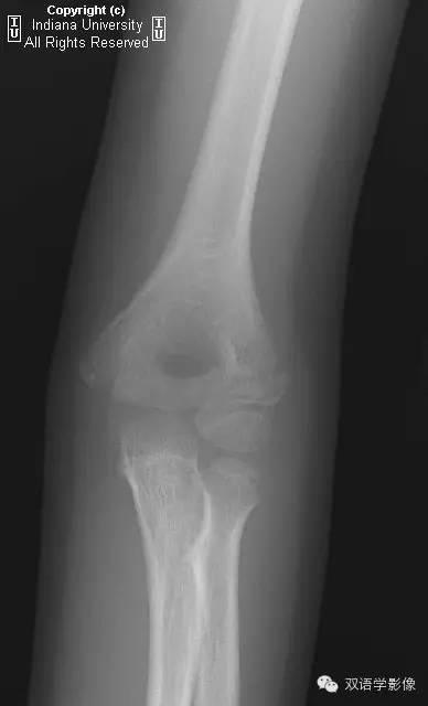
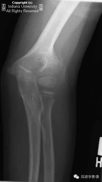
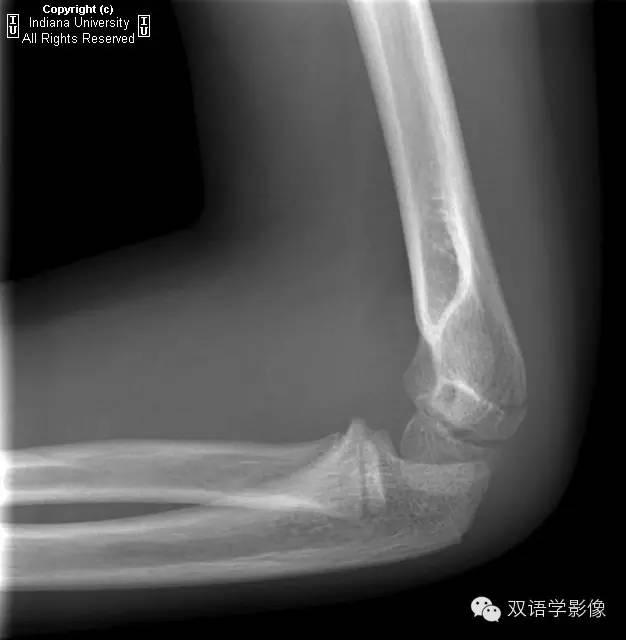
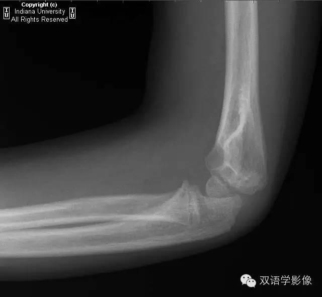
Here are the original images plus follow-up images at three and six weeks later.
下图为最初的X线片和三个月、六个月复查的X线片。
2.At what age does the lateral condyle typically ossify?
肱骨外髁骨化中心出现的年龄?
6
8
10
12
Never
3.Which category of Salter-Harris fracture is this considered to be?
此例骨骺损伤的Salter-Harris分型?
Type I
Type II
Type III
Type IV
选择题答案:
- Lateral condyle fracture
- 10
- Type IV
Findings
There is a left lateral condylar fracture, with anterior fat pad sign. Fragment is less than 2 mm displaced. Radial head is in place. Follow-up imaging reveals bony bridging compatible with healing.
肱骨外侧髁骨折伴前方脂肪垫征阳性。骨片移位小于2mm。桡骨小头在位。复查X线示骨痂形成、愈合良好。
Differential Diagnosis
Normal ossification center正常骨化中心Radial head dislocation桡骨小头脱位Medial condylar fracture内髁骨折Lateral condylar fracture外髁骨折
Diagnosis
Lateral condylar elbow fracture肘关节外髁骨折
Key Points
Lateral condylar elbow fracture is the second most common pediatric elbow fracture (15%).肘关节外髁骨折是儿童第二常见的肘部骨折(15%)。Seen most often from ages 4 to 10, with peak incidence at age 6.常见于4岁-10岁,发病高峰年龄为6岁。Usually related to a fall on an outstretched hand with the elbow extended and the forearm abducted.多于摔倒时手臂伸展有关,一般是前臂相对固定、肘关节过伸所致。Considered to be a Salter-Harris type IV injury.该损伤属于Salter-Harris IV型。Stage or type I fractures with less than 2 mm displacement can be treated with immobilization.移位小于2mm的I型损伤一般采取固定治疗。Open reduction recommended for all type II or III fractures.Salter-Harris II型或III型,推荐切开复位。Easily missed if thought to represent a normal ossification center. The lateral condyle does NOT ossify until age 10.如果误认为这是一个正常的骨化中心,则很可能漏诊该病。实际上,在10岁之前,这里不会出现骨化中心。
来源:双语学影像
,


免责声明:本文仅代表文章作者的个人观点,与本站无关。其原创性、真实性以及文中陈述文字和内容未经本站证实,对本文以及其中全部或者部分内容文字的真实性、完整性和原创性本站不作任何保证或承诺,请读者仅作参考,并自行核实相关内容。文章投诉邮箱:anhduc.ph@yahoo.com